
Digestive System: Part I
(Part II will cover the 4 glands: Salivary, Pancreas, Liver, & Gall bladder)
The digestive system is a system of connecting tubes in the body, from the mouth to the anus.


- Cells that make up digestive tract & their function
- How these cells, tissues, structures, & organs work in digestion
- Unique features of different organs, regions, & structures in the digestive system
- Pathology of structures
 |
| From slideplayer |
The outer layer is either a serosa or adventitia depending on whether it is touching anything or not.
- Serosa: Within the peritoneal cavity
- Adventitia: touching another organ in the posterior abdominal wall (retroperitoneal)
 |
| From BBC education |
- Mouth
- Lips (and cheeks)
- Thin skin part (cutaneous)
- Has hair follicles, sebaceous glands, sweat glands
- Colorized part (Vermillion border/part of the lip we see on the outside)
- Very vascular, will have Meissner's corpuscles
- Oral part
- The part that touches your teeth
- No more keratin
- Will have mucous glands
- Palates
- Hard Palate
- Stratified squamous with keratin
- Soft Palate
- Will have 2 epitheliums depending on which side: mouth or nose side
- Mouth side
- stratified squamous
- Nose side
- pseudostratified squamous with cilia & goblet cells
- Tonsils
- Teeth
- Odontoblasts make dentin
- Ameloblasts make enamel
- No way to regenerate more enamel
- Tongue
- Filiform Papillae
- General sense (sensory nerve)
- Anterior 2/3 tongue = cordi tympani innerv. this base
- Foliate Papillae
- Fungiform Pappilae
- Taste buds
- Circumvallate Papillae
- Form the line you see towards the back of your tongue
- Line marks the anterior 2/3 & posterior 1/3 of tongue
- Collects food particles (via collecting the saliva)
- Taste buds (see above)
- Salivary Glands
- Secrete saliva into mouth to wash away particulate matter so that you can go from one taste to another
- Esophagus
- Stomach
- Has rugae to allow stomach to stretch
- Has 3 parts
- Cardiac stomach
- Fundic stomach
- Pyloric stomach
- Small Intestine
- Lots of villi
- increases surface area for food absorption and adding digestive secretions
- Has 3 parts
- Duodenum
- (Jejunum--hard to tell)
- Ileum
- (Appendix)
- "Blind-ending sac"
- Will see crypts in the lamina propria
- Will see lymph nodules in the submucosae
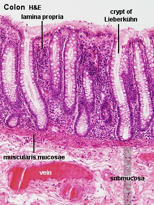
- Large Intestine (Colon)
- No villi
- For transporting waste, storing & absorbing water and ions, meaning lots of goblet cells
- Rectum
- Anal Canal (Anus)
 |
| Medical Faculty of Udayana University |
 |
| Slideshare |
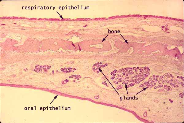 |
| Soft Palate, Southern Illinois University |
 |
| Michigan Medical School |
 |
| Steph Sadler |
 |
| From Colostate.edu |
| Esophagus histopathology moment:Below is a normal esophagus Potential histopathology of esophagus: Barrett's esophagus (dysplasia of epithelium) 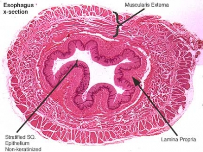 |
| faculty.une.edu |
Stomach: Fundic vs. Pyloric Fundic: short pits, long glands Pyloric: long pits, short glands |
 |

Small Intestine:
Duodenum, Jejunum, & Ileum
 |
| University of Western Australia |
 |
| University of Western Australia |

 |
| University of Western Australia |
 |
| University of Western Australia |
 |
| University of Western Australia |


Nice post. Well what can I say is that these is an interesting and very informative topic on food bad for digestion
ReplyDelete